At the end of this chapter, you will be able to:
- identify the different parts of the respiratory system
- explain how your child breathes
- understand how a tracheostomy helps your child breathe
A child’s respiratory system (breathing system) can be broken down into the:
- upper respiratory tract
- lower respiratory tract
Upper respiratory tract
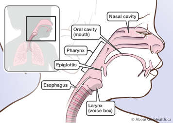
Nasal cavity
When you breathe through your nose, the air is warmed, moisturized, and cleaned. Tiny hairs called cilia line the inside of the nose and filter the air.
Oral cavity (mouth)
When you breathe through your mouth, you are warming and moisturizing the air. However, there are no cilia in the oral cavity, so the air is not filtered.
Pharynx
The pharynx is a muscular tube between the nose and the mouth.
Larynx (voice box)
The larynx (voice box) is between the pharynx and the trachea. It contains the vocal cords. When air is breathed in and out, voice sounds are created here. The vocal cords can be closed to build up pressure in the lungs and create a strong cough.
Epiglottis
The epiglottis is a flap that hangs over the larynx. When you swallow, this flap covers the larynx so food and/or drink will go into the esophagus and not into the trachea and lungs.
Esophagus
The esophagus is the feeding tube that connects the pharynx and the stomach.
Lower respiratory tract
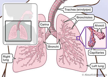
Trachea (windpipe)
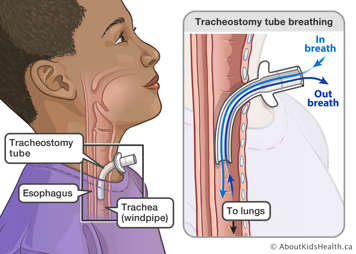
The trachea is the breathing tube that connects the larynx to the lungs. This is where a tracheostomy tube is inserted.
Lungs (right and left)
The lungs are the two organs used for breathing in the body. The lungs take in oxygen from the air and release carbon dioxide.
Carina
The carina is where the trachea divides into two hollow tubes, called the right and left bronchi.
Bronchi and bronchioles
The bronchi are tubes that supply air to each lung. The bronchi divide into smaller and smaller hollow tubes called bronchioles. These are the smallest air tubes in the lungs.
Alveoli
The alveoli are tiny sac-like structures at the tip of the bronchioles. They allow oxygen and carbon dioxide to move in and out of the lungs.
Pleura
The pleura are membranes that surround the lungs. The visceral pleura is the inside membrane, attached to the lungs. The parietal pleura is the outside membrane.
Capillaries
The capillaries are blood vessels in the walls of the alveoli.
Ribs
The ribs are the individual curved bones that surround the lungs.
Ribcage
The ribcage is the group of ribs that protect the lungs.
Respiratory muscles
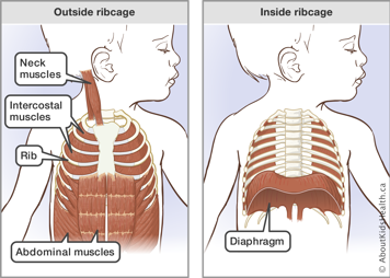
The respiratory muscles play a vital role in every breath a child takes. They include the:
- diaphragm
- neck muscles
- intercostal muscles
- abdominal muscles
Diaphragm
The diaphragm is a large, sheet-like muscle. The diaphragm is the main muscle involved in breathing. It is always active.
Neck muscles
If a child is having difficulty breathing, the muscles of the neck can help.
Intercostal muscles
The intercostal muscles are the muscles between the ribs that help with breathing in and out.
Abdominal muscles
The abdominal muscles are active during a forced exhalation (breathing out) only. They also help to create a good cough.
Breathing
Why do we breathe?
All living things need a constant supply of oxygen to live. Our respiratory system takes this oxygen from the air, and gets rid of a waste gas called carbon dioxide. The process of breathing usually takes place without us thinking much about it.
Breathing is a two-stage process:
- inhalation (breathing in)
- exhalation (breathing out)
When we breathe in, oxygen enters the lungs. When we breathe out, carbon dioxide exits the lungs.
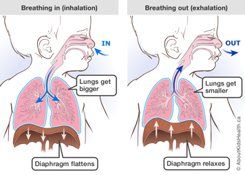
Breathing in
During inhalation, the large diaphragm muscle moves down and out, pulling air into the lungs. In addition, the intercostal muscles pull the ribcage upwards and outwards.
Air enters through the nose or mouth and passes into the trachea, bronchi, bronchioles and finally into the alveoli. In the alveoli, oxygen breathed in from the air is transferred into the capillaries. It is then pumped by the heart all over the body to deliver oxygen.
Breathing out
During exhalation, the lungs get smaller as the diaphragm relaxes and moves back. The intercostal muscles pull the ribcage downwards and inwards (towards the spine).
Carbon dioxide, the waste gas of the lung, moves from the capillaries to the alveoli. It then passes into the bronchioles, bronchi and trachea before leaving the body through the nose and mouth. The lungs, like a stretched spring, recoil back to their unstretched state and the air is squeezed out.
This process of breathing usually takes place without you thinking much about it. The diaphragm and intercostal muscles are usually relaxed when you breathe out. However, you can contract these muscles to push air out quickly, for example when you cough or sneeze.
Breathing with a tracheostomy
Your child breathes in and out through a tracheostomy (trach) tube. This is a hole through the skin in the front of the neck and into the trachea. The hole is created during surgery. The opening in the neck is called a stoma.
With tracheostomy tubes, the air goes directly into the lungs and does not pass through the nose or mouth first. Depending on your child’s size and type of tracheostomy tube, some air may still go in and out of your child’s nose or mouth.
Since the air bypasses the nose, it must be warmed, moistened and filtered before going into the tracheostomy tube. This can be done in different ways, such as humidifiers or a heat moisture exchanger. This will be discussed in future chapters where you will learn more about a tracheostomy tube and management of your child’s artificial airway.
