Your premature baby needs an eye exam by a paediatric ophthalmologist. This person is an eye doctor who specializes in checking and treating eye problems in children, including retina problems. This exam is important because premature babies may develop a condition called retinopathy of prematurity, or ROP.
What is the retina?
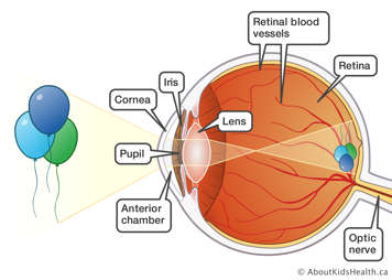
The retina is the inside lining in the back of the eye. It is the part that absorbs the rays of light that enter the eye. The retina changes the rays into electrical signals. It then sends the signals to the brain as a picture. In this way, the retina is like a film in a camera: it turns the rays of light it receives into a picture that a person sees.

What is retinopathy of prematurity?
Retinopathy of prematurity (ROP) happens in premature babies when abnormal blood vessels start developing at the back of the eye.
At about 16 weeks of pregnancy, the fetus’s retina begins to develop blood vessels. The blood vessels that feed the retina start at the back of the eye and grow toward the front. The retina is gradually and evenly covered in blood vessels. They finish forming just before the baby is born at full term.
In a premature baby, these blood vessels have not finished forming. They continue to form after the baby is born. Most of the time, they will form normally and there will be no problem. But if smaller, abnormal vessels start developing, the condition is called ROP.

What can happen if a baby has ROP
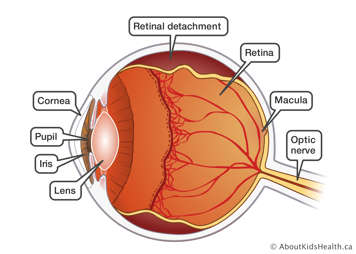
Abnormal vessels can lead to bleeding and scarring in the retina. They may also cause the retina to separate, or move, from its normal place in the eye. This is called retinal detachment. This could lead to poor vision or even blindness.

How common is ROP?
Nobody really knows why abnormal blood vessels form. Some premature babies need extra oxygen to help them breathe. It is thought this oxygen treatment may play a part in ROP, even when it is closely monitored.
Not all premature babies have ROP. Babies born before 28 weeks or who weigh less than 1000 g at birth are at the highest risk for developing severe ROP. The risk of ROP developing also depends on how well the retina has formed.
Checking your baby for ROP
In Canada, all premature babies with a birth weight of 1250 g or less, or who are born at or before 30 weeks and six days are routinely examined for ROP. They will likely be examined initially at four to six weeks after birth.
The ophthalmologist will check your baby's eyes for any abnormal vessels. If these vessels are treated in time, it may help to stop retinal detachment.
Here is what you can expect to happen during the exam.
- Your baby will have special eye drops to make the pupils bigger. The pupil is the dark area in the centre of the coloured part of the eye. The drops take 30 minutes to an hour to work, sometimes longer.
- Since your baby needs to be very still when their eyes are checked, they will be wrapped in a blanket and held down gently.
- The doctor will check the retina using an instrument with a bright light called an ophthalmoscope.
- Your baby will have eye drops to numb the surface of the eyeball.
- Once the eyeball is numb, the doctor will use an instrument called a speculum to hold your baby's eyelids apart. This is because your baby is too young to keep their eyes open.
- To get a good look at the eye, the doctor will also use an instrument called a depressor to gently move the eyeball.
- Sometimes, a photograph will be taken of the retina using a special camera.
Your baby should not feel any pain.
Being held down and having a bright light shone in their eyes will make your baby uncomfortable. They may cry during the exam, but they should not feel any pain.
Explaining the condition of your baby's eyes
After the exam, the doctor will explain the condition of your baby's eyes. Your baby's condition will be graded depending on how much the abnormal blood vessels have grown. The doctor will use the terms "zone", “type” and "stage".
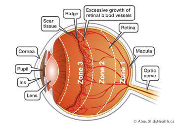
- The zone is graded from 1 to 3. This explains how far the blood vessels have grown on the retina. Vessels go from Zone 1 to 3 as they grow. The second image on this page shows the zones.
- The stage explains the severity of the ROP. It is graded from 1 to 5. Stage 1 is the best (least severe) and Stage 5 is the worst (most severe).
-
Stage 1 ROP
In Stage 1, there is a thin line between the area with blood vessels and the area where the blood vessels have not grown yet. At this stage, the vessels may grow normally on their own, but the condition must be watched.
-
Stage 2 ROP
In Stage 2, the line between the areas with and without blood vessels widens and thickens into a ridge. The condition may still resolve, or it may progress to Stage 3 ROP.
-
Stage 3 ROP
In Stage 3, new blood vessels start to grow along the ridge and extend into the clear gel that fills the eye, called vitreous body. These blood vessels can bleed and form scar tissue.
-
Stage 4A ROP
In Stage 4A, the abnormal blood vessels and scar tissue pull on the retina, partially detaching it. The centre of vision, called fovea, is not involved.
-
Stage 4B ROP
The retina is still only partially detached, but the fovea is affected. This usually leaves both the centre and peripheral vision impaired to some degree.
-
Stage 5 ROP
The retina is completely detached, severely affecting vision.
- The terms ‘type 1’ and ‘type 2’ ROP are descriptions that are used to identify babies whose eyes show changes of ROP that require treatment (type 1) or show changes that do not require treatment but must be carefully monitored (type 2).
How often your baby's eyes will be checked
Your baby's eyes need to be checked often. Sometimes they will be checked every week or even every few days. The frequency of exams depends on different factors, such as the severity and location of ROP in the eye and how quickly normal blood vessels are forming. The formation of blood vessels is called vascularization. After each exam, the doctor will let you know when the next exam will happen.
In the majority of cases, even when ROP develops, it will resolve on its own with minimal effect on your baby’s vision. However, it is very important to keep all the appointments because abnormal vessels can form fairly quickly. In approximately 5 per cent of babies, ROP will progress to the extent that is no longer safe to wait for it to resolve on its own.
Treating ROP
If your baby has ROP, the treatment will depend on your baby's eye condition. Your baby may need laser treatment, eye injections or even surgeries (operations) to repair retinal detachment. Your baby’s ophthalmologist will discuss the best treatment for your baby with you.
Caring for your child's eyes in the future
All premature babies need regular eye exams by an ophthalmologist, preferably a paediatric ophthalmologist.
If your baby's retinal blood vessels are normal
Your baby will still need to see an ophthalmologist regularly, but they will no longer need a retina specialist. Your child’s health-care provider may refer your child to a paediatric ophthalmologist in your area for check-ups.
If your baby has been treated for ROP
Your baby will continue to see a paediatric ophthalmologist who also specializes in retinal problems.
When your baby's retinal condition is stable, their health-care provider may then refer them to a paediatric ophthalmologist in your area for continued check-ups.
Regular check-ups are important
It is important that your child is seen by an ophthalmologist often. Even when the blood vessels are fully formed, there is a greater chance that a premature baby can have certain eye conditions in the future, such as:
- near-sightedness (myopia)
- cross eyes (strabismus)
- lazy eye (amblyopia)
- a condition in which the rays of light are focused in each eye at a different point (anisometropia)
The ophthalmologist will want to check your child's eyes for these conditions so that they can be treated.
